In vivo optical imaging serves as a cornerstone of biomedical research by offering insights into complex physiological and pathological processes. However, the immense potential of the emission window beyond 1500 nm which combines the advantages of deep penetration and minimized background interference has long been severely constrained by the lack of effective molecular sensing probes. To overcome this critical bottleneck, the research team introduces an electronic-symmetry-tuned emission (ESTE) mechanism in monofunctionalized Er(III)-phthalocyanine complexes, establishing a general molecular platform in vivo dynamic chemical sensing beyond 1500 nm.
The team first synthesized monofunctionalized Erbium(III)-phthalocyanine complexes and discovered that the electronic effects of peripheral substituents disrupt the inherent symmetry of the molecular structure. Significantly, this molecular-level "symmetry breaking" did not dampen the characteristic 1530 nm near-infrared emission of Erbium(III); instead, it precisely reshaped the orbital distribution of the ligands, thereby enabling flexible tuning of the excitation profiles. Owing to efficient triplet energy transfer from the phthalocyanine ligands to the central Erbium(III) ion via the antenna effect, the complexes maintained bright 1530 nm emission while exhibiting highly tunable excitation wavelengths (from 680 nm to 705 nm). This innovative design enabled the construction of a dual-excitation ratiometric signal mode, whereby monitoring the ratio of 1530 nm emission intensities under two distinct excitation wavelengths achieves high-precision quantitative detection independent of probe concentration, providing a core tool for dynamic in vivo imaging.
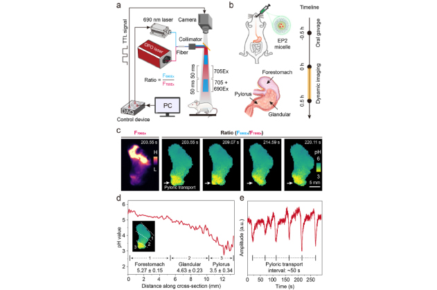
Figure 1. (a) Schematic of the dual-excitation dynamic imaging setup.
(b) Schematic of the gastric imaging protocol using EP2 micelles. The inset shows mouse stomach anatomy.
(c) Dynamic intensity (F705Ex) and ratiometric (F690Ex/F705Ex) imaging of the mouse stomach.
(d) Cross-sectional pH distribution in the fed mouse stomach.
(e) Pyloric transport dynamics.
Leveraging this strategy, the team developed a pH-responsive probe (EP2) encapsulated in micelles, enabling real-time imaging of dynamic physiological processes in the mouse stomach. Given the complex anatomical structure of the stomach (forestomach, glandular stomach, and pylorus), conventional intensity-based imaging is restricted by uneven probe distribution and fails to provide accurate quantification. In contrast, ratiometric NIR-II-L fluorescence imaging successfully revealed distinct pH spatial gradients in the stomachs of feeding mice, where the pyloric region exhibited significantly higher acidity compared to the forestomach. Compared to traditional invasive assessments requiring hours or even days, this non-invasive real-time imaging approach substantially advances the ability to elucidate gastrointestinal physiological dynamics.
The probe exhibited a rapid ratiometric fluorescence response to local Cu(II) concentration changes within 10 s of injection, and clearly delineated the fan-shaped diffusion dynamics of Cu(II) within the abdominal cavity in approximately 150 s. Benefiting from the exceptionally low tissue background interference at 1530 nm, the team clearly resolved the complex anatomical contours of the mouse intestine and successfully recorded intestinal peristaltic movements at a frequency of 0.46 Hz. This enables not only dynamic tracking of metal ions, but also offers a novel visualization strategy for non-invasive assessment of physiological mechanical functions, such as intestinal motility.
The ESTE strategy proposed in this study eliminates the need for complex nano-assembly or high-power excitation; instead, it achieves precise tuning of excitation profiles solely through molecular-level structural design, offering a universal strategy for designing functional probes for deep-tissue sensing. This novel single-molecule complex-based sensing platform holds great promise for frontier applications such as tumor microenvironment analysis, neurotransmitter monitoring, and pharmacokinetic evaluation, paving the way for future high-resolution, non-invasive in vivo molecular diagnostics.
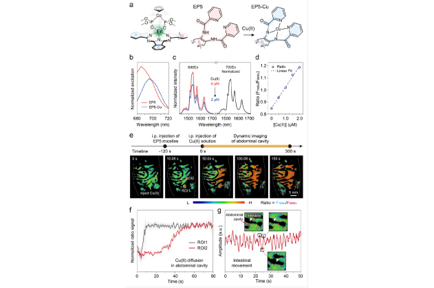
Figure 2. (a) Cu(II)-responsive mechanism of EP5.
(b) Normalized excitation spectra of EP5 and EP5-Cu.
(c) Cu(II)-dependent normalized emission spectra under 690 and 700 nm excitation. EP5: 5 μM.
(d) Linear excitation-ratiometric response to Cu(II) (0−2 μM).
(e) Top: schematic of the Cu(II) sensing protocol using EP5 micelles. Bottom: dynamic ratiometric imaging of Cu(II) diffusion in the mouse abdominal cavity.
(f) Time-dependent Cu(II) dynamics at ROI1 and ROI2.
(g) Intestinal movement dynamics.
References:
B. Wu, L. Zhang, K. Yan, M. Mei, W. Wu, Z. He, S. Wang, F. Zhang, Electronic-symmetry-tuned emission beyond 1500 nm in erbium(III)-phthalocyanine complexes for high-resolution in vivo biosensing. J. Am. Chem. Soc. 2025, 147, 44185–44190. https://doi.org/10.1021/jacs.5c13688

 xusy@digi-united.com
 +86 178 9511 3630


Design by DOHARD TEL: 0574 8808 1302
Copyright © 2023上海数联生物科技有限公司版权所有 | 沪ICP备2021012949号-1

